| Adineta gracilis var. 11, specimen from (1) |
| |
 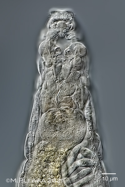 |
| Adineta gracilis var. 11, two images from anterior part of the same specimen. Left: ventral side of the head; focus plane on the ciliary field with V-shaped prehensile apparatus. In the trunk some parasites are visible. Right: slightly different focus plane; rostrum lamella with sensory cilia; the ciliary field is retracted and the head ring muscle is contracted. Clia of the buccal field and the buccal tube are also visible. |
| |
 |
| Adineta gracilis var. 11, integument of the lower lip and prehensile apparatus of a macerated specimen. |
| |
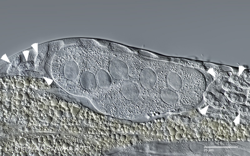 |
| Adineta gracilis var. 11. Up to now all A. gracilis-specimens that I observed had 4 nuclei in the vitellarium. This is the first population where specimens with 8 nuclei could be observed. Also visible in this specimen are fungal parasites (some are marked by arrowheads). (1) |
| |
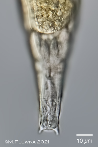 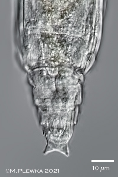 |
| Adineta gracilis var. 11, two aspects of the foot while creeping, different specimens. Left: extended specimen, right: slightly contracted specimen |
| |
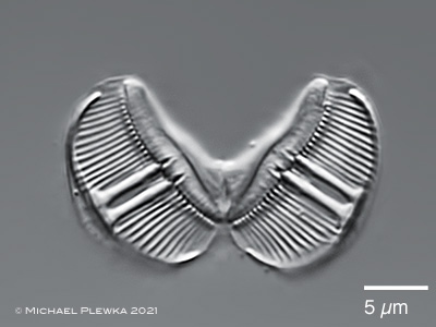  |
| Adineta gracilis var. 11, ramate trophi: Left: cephalic view; DF: 2/2. Righjt: caudal view, ramus length (raL): 13µm. |
| |
| >>>>>> see also Adineta gracilis |
| |
| |
|
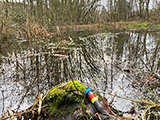
Location (1): Sprockhoevel Schee, NRW, Germany, pond |
 |
| |
| Habitat (1): detritus between desintegrating leafs |
| |
| Date (1): coll.: 30.12.2021; img.: 31.12.2021 |
| |
| |
|
|
|
|
|
| |
|
| |
|
|
| |
| |